
Most cancers is a extremely complicated illness, partly due to the ecosystems (or microenvironments) through which tumors reside. With a large number of cell sorts and microorganisms, the tumor microenvironment can affect how most cancers kinds, whether or not it spreads, and the way it responds to therapy. Making progress towards most cancers, subsequently, would require an intensive understanding of the intricate relationships that exist between cancers and their microenvironments.
Luckily, latest technological advances have enabled researchers to check the tumor microenvironment in unprecedented element and decipher a lot of its complexities.
A plenary session on the American Affiliation for Most cancers Analysis (AACR) Annual Assembly 2024, titled “Profiling Tumor Ecosystems in Native Tissue Context,” spotlighted a few of the revolutionary approaches used to look at the tumor microenvironment, in addition to the discoveries these approaches have yielded to this point.
“Most cancers is exceedingly complicated, and it has taken the event of applied sciences that may perceive most cancers at this stage of complexity to essentially begin making in-roads,” mentioned session moderator Christine Iacobuzio-Donahue, MD, PhD, a researcher at Memorial Sloan Kettering Most cancers Middle and the Editor-in-Chief of the AACR journal Most cancers Analysis.
“I firmly consider we’re nicely on our solution to beating most cancers due to … most of these approaches.”
Convergence of A number of Applied sciences Supplies Perception Into Distinct Cancers
Michael Angelo, MD, PhD, of Stanford College College of Medication, shared a novel framework for finding out spatial tumor biology, which he and colleagues used to grasp illness development and therapy in a number of most cancers sorts.
Their multimodal framework depends on three strategies to characterize elements of the tumor microenvironment: multiplexed ion beam imaging by time of flight (MIBI-TOF) to map proteins, spatial transcriptomics to map gene expression, and matrix-assisted laser desorption/ionization (MALDI) to map glycan sugars within the tissue. They then make use of machine studying to research the information at three spatial scales: the pixel stage, the entire cell stage, and the multicellular neighborhood stage.
Angelo and colleagues utilized their framework to characterize the expression patterns of seven frequent drug targets in glioblastoma. In doing so, the researchers uncovered alternatives to enhance therapy efficacy for this extremely deadly illness.
They discovered that after they handled tumors with two focused remedy medication as an alternative of 1, they had been in a position to goal about 20% extra most cancers cells. Growing to a few or extra medication, nevertheless, solely marginally impacted the variety of cells focused. This was as a result of a big portion of glioblastoma cells didn’t specific any of the seven druggable targets.
Based mostly on these findings, Angelo proposed that therapy for glioblastoma would possibly profit from a two-pronged technique that mixes dual-targeted remedy with interruption of immune suppression to allow clearance of most cancers cells that lack druggable targets. To disrupt immune suppression, Angelo recommended concentrating on sialic acid-modified glycans, which he and colleagues discovered to be enriched in superior glioblastomas.
Angelo and colleagues additionally utilized their multimodal strategy to establish spatial markers of illness development in ductal carcinoma in situ (DCIS), a noninvasive type of breast most cancers. By evaluating tissue from sufferers whose DCIS did or didn’t progress to invasive breast most cancers, the researchers recognized a number of components that elevated the chance of development.
These included modifications to the spatial group of 14 completely different cell populations, disruptions to the myoepithelium (a concentric tissue layer that separates DCIS cells from the encircling stroma), and altered ductal topology. Once they mixed these determinants right into a danger prediction profile, they had been in a position to predict 10-year DCIS development with an accuracy of 82%, Angelo reported.
Lastly, he and colleagues analyzed metastatic triple-negative breast tumors to establish spatial options that would predict response to nivolumab and chemotherapy, which they used to develop a danger prediction mannequin for illness relapse. The objective of such a mannequin can be to establish sufferers who’re probably to learn from the routine earlier than they start therapy and keep away from pointless therapy for these much less more likely to profit.
Nonetheless, the researchers discovered that, whereas their mannequin precisely predicted relapse for sufferers already present process therapy, it had restricted accuracy previous to therapy. These findings recommend that spatial determinants of response, similar to these recognized right here, can change over the course of remedy, Angelo famous.
Regional Variation of Intratumoral Microbes Impacts Most cancers Cell Features
Microbes within the human physique can have optimistic and unfavorable impacts on human well being. Within the session’s second presentation, Susan Bullman, PhD, of the Fred Hutchinson Most cancers Middle, mentioned how these microbes can influence most cancers, sharing that the spatial distribution of microbes inside tumors impacts mobile features.
She confirmed that Fusobacterium nucleatum had been heterogeneously distributed throughout gastrointestinal tumors and that tumor areas enriched for the micro organism had been immune suppressed and necrotic. Since necrosis ends in decreased oxygen ranges, necrotic areas are nicely fitted to anaerobic micro organism similar to F. nucleatum, Bullman famous.

Utilizing spatial transcriptomics and a newly developed methodology known as INVADEseq, Bullman and colleagues discovered that the presence of micro organism altered gene expression of cells within the tumor microenvironment. For instance, F. nucleatum induced upregulation of mobile pathways related to cell migration and metastasis in gastrointestinal cancers.
Bullman and colleagues additionally used microscopy strategies coupled with single-cell transcriptomics to concurrently visualize bacterial populations and gene expression modifications throughout colon cancers.
“Certainly, these intratumoral microbes are actively modulating their contaminated tumor area of interest inside the tumor microenvironment,” she famous. She added that understanding how microbes influence most cancers development and therapy response could allow researchers to intercept unfavorable host-microbial interactions and enhance affected person outcomes.
Multimodal Spatial Transcriptomics to Perceive Most cancers Evolution
Joakim Lundeberg, PhD, of KTH Royal Institute of Know-how and Science for Life Laboratory, mentioned the ability of mixing spatial transcriptomics with different modalities to raised perceive most cancers initiation and development.
Early functions of spatial transcriptomics revealed the huge heterogeneity inside most cancers tissue, and mixing typical spatial transcriptomics with single-cell RNA sequencing offers even higher perception into the heterogeneous gene expression and cell sorts discovered within the tumor microenvironment, Lundeberg defined.

He and colleagues utilized this strategy to map the modifications that happen as precancerous situations evolve to prostate or breast most cancers. They inferred gene copy quantity modifications from gene expression knowledge, with greater and decrease gene expression doubtlessly indicative of chromosome positive factors and losses, respectively.
They discovered that the frequency of inferred copy quantity modifications elevated as regular prostate tissue advanced to premalignant prostatic intraepithelial neoplasia (PIN) after which to prostate most cancers. Additional, they found that cells harboring these copy quantity modifications had been localized to glands.
In breast most cancers tissue, Lundeberg and colleagues noticed distinct inferred copy quantity modifications when evaluating noninvasive in situ breast most cancers clones with invasive clones, with positive factors in chromosome 2 and chromosome 19, respectively.
Lundeberg additionally mentioned the potential of spatial multiomics, which mixes knowledge on the spatial distribution of gene expression, metabolites, lipids, glycans, peptides, and histology, to establish biomarkers of most cancers development.
Lundeberg and colleagues are at the moment utilizing deep studying to compile spatial multiomics knowledge from tissue sections into three-dimensional tissue fashions to raised perceive the modifications that happen throughout tumor tissues.
Including a New Dimension to Finding out Melanoma
Whereas tissue profiling is conventionally carried out on skinny slices of tissue (sometimes 5 microns thick), Peter Sorger, PhD, of Harvard Medical College, argued that this strategy is main researchers to overlook key info. Skinny slices don’t seize entire cells and even entire nuclei, he defined, so thicker sections are wanted to correctly characterize tissue pathology.
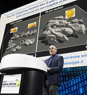
Sorger shared outcomes from a newly developed methodology for the molecular profiling of tissue slices as much as 35 microns in thickness. This allowed the researchers to look at cells throughout all three dimensions of melanoma tissues (size, width, and depth), which uncovered that the assorted cell populations that made up the tissue had been densely packed and infrequently intertwined—options that weren’t detected by typical two-dimensional approaches.
An evaluation of early melanoma tissue revealed heterogeneous expression of assorted immune markers, with inflammatory markers localized to websites the place the melanoma had began to invade surrounding tissue. Additional evaluation confirmed that metastatic melanomas had equally heterogeneous gene expression, which was organized round tissue vasculature, perinecrotic domains, and tumor margins.
Two-dimensional and three-dimensional imaging each have professionals and cons, Sorger famous. Two-dimensional imaging has restricted decision however is easier and might subsequently be used to research numerous specimens. In distinction, three-dimensional imaging offers deep, useful characterization however of a comparatively small variety of samples. Sorger hopes to discover a solution to mix one of the best of each strategies.
“We actually now are pushing onerous to … develop machine studying algorithms to drag these worlds collectively,” he mentioned, noting that performing analyses with the depth of three-dimensional imaging however with the benefit of two-dimensional imaging would drastically enhance single-cell analyses.


